
Lahabali kɔligu:Echinococcus Life Cycle.png
Echinococcus_Life_Cycle.png (600 × 571 pixels, file size: 44 KB, MIME type: image/png)
Lahabali jia
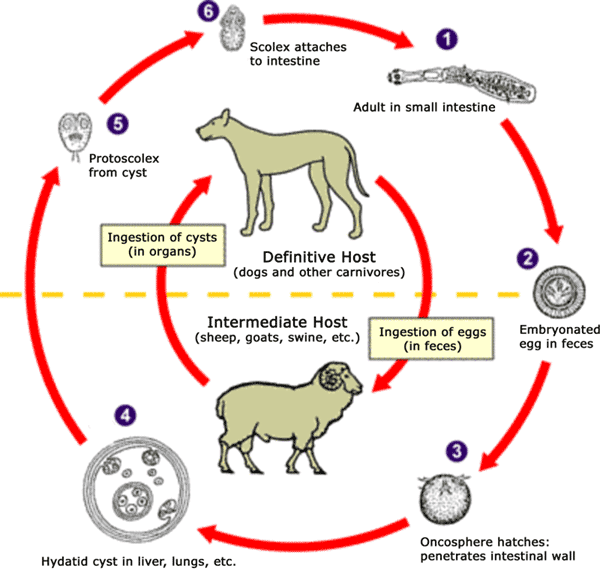
The adult Echinococcus granulosus (3 to 6 mm long) [1] resides in the small bowel of the definitive hosts (dogs or other carnivores). Gravid proglottids release eggs [2] that are passed in the feces. After ingestion by a suitable intermediate host (under natural conditions: sheep, goat, swine, cattle, horses, camel), the egg hatches in the small bowel and releases an oncosphere [3] that penetrates the intestinal wall and migrates through the circulatory system into various organs, especially the liver and lungs. In these organs, the oncosphere develops into a cyst [4] that enlarges gradually, producing protoscolices and daughter cysts that fill the cyst interior. The definitive host becomes infected by ingesting the cyst-containing organs of the infected intermediate host. After ingestion, the protoscolices [5] evaginate, attach to the intestinal mucosa [6] and develop into adult stages [1] in 32 to 80 days. The same life cycle occurs with E. multilocularis (1.2 to 3.7 mm), with the following differences: the definitive hosts are foxes, and to a lesser extent dogs, cats, coyotes and wolves; the intermediate host are small rodents; and larval growth (in the liver) remains indefinitely in the proliferative stage, resulting in invasion of the surrounding tissues. With E. vogeli (up to 5.6 mm long), the definitive hosts are bush dogs and dogs; the intermediate hosts are rodents; and the larval stage (in the liver, lungs and other organs) develops both externally and internally, resulting in multiple vesicles. E. oligarthrus (up to 2.9 mm long) has a life cycle that involves wild felids as definitive hosts and rodents as intermediate hosts. Humans become infected by ingesting eggs , with resulting release of oncospheres in the intestine and the development of cysts in various organs.
Image adapted from original available at the United States Centres for Disease Control Parasitology Identification Laboratory ([1] archive copy at the Wayback Machine).

|
File:Echinococcus Life Cycle.svg is a vector version of this file. It should be used in place of this PNG file when not inferior.
File:Echinococcus Life Cycle.png → File:Echinococcus Life Cycle.svg
For more information, see Help:SVG. |
 |
Fukumisi shahira niŋbu
This image is a work of the Centers for Disease Control and Prevention, part of the United States Department of Health and Human Services, taken or made as part of an employee's official duties. As a work of the U.S. federal government, the image is in the public domain.
|
Faal tarihi
Dihimi dabisili/saha n-nya kɔl' bihi kamani di ni di yina shɛm
| Zuŋɔ dabisili/Saha | Thumbnail | Di tarisi | ŋun su | tɔhibu | |
|---|---|---|---|---|---|
| din na chana | 10:54, 24 Silimin gɔli January 2007 | | 600 × 571 (44 KB) | Pngbot | optimized with optipng |
| 01:48, 26 Silimin gɔli April 2005 |  | 600 × 571 (55 KB) | FirstPrinciples~commonswiki | Smaller & clearer | |
| 01:36, 26 Silimin gɔli April 2005 |  | 800 × 761 (79 KB) | FirstPrinciples~commonswiki |
Lahibali kɔligu zaŋ tum tuma
Yaɣi shɛli kani din mali lahabali kɔligu ŋɔ n-kuri bukaata.